When Katie Couric revealed her breast cancer diagnosis, many women were surprised that her lump was discovered during an ultrasound exam, an imaging test performed in addition to a mammogram for women with dense breasts.
Dense breasts are common—nearly half of all women have them. But while many women know they have dense breasts (federal law now mandates that all women are informed of their breast density), many don’t know that having dense breasts comes with a slightly elevated risk of developing breast cancer and that they could benefit from additional imaging tests.
What are dense breasts?
Breast density has nothing to do with firmness or size, says Lauren Friedlander, MD, assistant professor of radiology at Columbia University Vagelos College of Physicians and Surgeons.
“Dense breasts are not something that you can notice or that your doctor can feel on physical examination,” she says. “It's a designation purely defined by what your mammogram pictures look like.”
Breasts are a mixture of fatty tissue and fibroglandular tissue. Every woman has a different mixture of these two types, but women who have more of the fibroglandular tissue are considered to have dense breasts. Breast density can decline over time, so women who have dense breasts in their 40s may not have dense breasts in their 60s.
In cancer screening, dense breasts are an issue, “because fibroglandular tissue appears white in a mammogram, just like many of the malignancies we look for,” Friedlander says. “And that means that finding malignances can be more of a challenge in women with dense breasts.”
3D mammography may improve detection
For women with dense breasts, an imaging exam called breast tomosynthesis, or 3D mammography, can help radiologists see cancers more clearly.
The experience of getting a 3D mammogram is essentially the same as conventional mammography from the patient’s point of view, but the machine gives the radiologist a more detailed look at the breast. 3D mammography does not create a true 3D image but produces multiple images, essentially dividing breast tissue into many thin slices.
With a stack of images, the radiologist can examine the breast layer by layer. “Things that might be hidden in a standard two-dimensional image are uncovered in breast tomosynthesis,” Friedlander says, “and we find a few more breast cancers that way.”
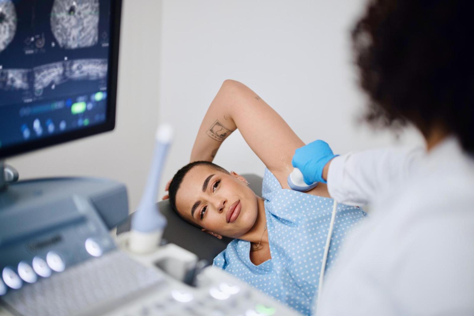
Extra screening with ultrasound
Any woman who has dense breasts may want to consider supplemental screening, usually with breast ultrasound.
Studies show that screening with ultrasound, in addition to mammography, improves detection of breast cancers in women with dense breasts.
A breast ultrasound is performed by an ultrasound technologist with a handheld probe. The technologist applies lubricating gel to the breast and moves the probe across the breast. The breasts are not compressed as they are during a mammogram, although the probe is applied with a little pressure.
One downside of additional screening tests is that women will get more false positives, Friedlander warns. “Particularly for a woman who comes for her first ultrasound, we may find lots of new things, but most are not cancer,” she says. Women who have ultrasound screening are more likely to need a biopsy and short-term follow-up for something that ultimately is benign.
Friedlander says that for some women with dense breasts, sticking to mammography and forgoing additional imaging is a reasonable option.
“There’s no one-size-fits-all plan anymore. And frankly, mammography is the only test that's proven to have a survival benefit,” she says. “In an ideal world, every patient would have a discussion with her physician and make these decisions together.”
Extra screening with MRI
Breast MRI takes screening one step further and is more sensitive, detecting even more cancers than ultrasound.
A breast MRI is more time-consuming and requires an intravenous injection of a contrast agent, which comes with a small risk of an allergic reaction. And as with ultrasound, patients screened with MRI are more likely to need biopsies for benign lumps.
“For these reasons, we are careful about who we bring in for breast MRI,” Friedlander says. “We do not use it for average-risk women who have dense breasts but reserve it for women who are in a higher risk category.”
Don’t put off mammography

Though dense breasts many pose a challenge when it comes to cancer detection, Friedlander says that regular mammography is still important.
Mammography is particularly good at revealing tiny calcifications or mineral deposits in the breast, which don't show up on ultrasound. “These calcifications can be the earliest signs of breast cancer,” Friedlander says, “so even women who are having MRI and ultrasound benefit from having a mammogram.”
“Having dense breast tissue does not mean that you will get breast cancer, but it does mean that you need to be vigilant in getting mammograms once a year, every year, and adding whatever supplemental screening test is appropriate for your level of risk.”
"breast" - Google News
October 14, 2022 at 11:48PM
https://ift.tt/ARVuU7D
Dense Breasts Explained - Columbia University Irving Medical Center
"breast" - Google News
https://ift.tt/6e5ISrB
https://ift.tt/od6k7yf
Bagikan Berita Ini


0 Response to "Dense Breasts Explained - Columbia University Irving Medical Center"
Post a Comment